2013 Annual Science Report
 University of Illinois at Urbana-Champaign
Reporting | SEP 2012 – AUG 2013
University of Illinois at Urbana-Champaign
Reporting | SEP 2012 – AUG 2013
Culturing Microbial Communities in Controlled Stress Micro-Environments
Project Summary
In NAI Theme 4B, our goal in Year 1 has been to initiate our understanding of how cells structure their genomes in response to specific environmental stresses and to determine whether or not such mechanisms have been a major force in directing the evolution of cells in natural environments over evolutionary time. Natural environments are typically rather heterogeneous at small scales, as established by sampling from geothermal hot spring communities, and so it is important to understand the generic impact on the evolution and structure of microbial communities. Our first step towards probing this phenomenon has been to culture living bacterial populations within a small specially constructed microfluidic device (called the GeoBioCell), where strong physical, chemical and biological gradients can be imposed under carefully controlled conditions.
Project Progress
Two major steps have been completed in Year 1 toward the accomplishment of our research goals. The first is that the Archaea Methanosaricina acetivorans, was chosen as a key lab organism on which to test microbial responses in our GeoBioCell microfluidic cells. The project team has worked extensively with this strain of Archaea, and its full genome has been sequenced to completion. Initial successful steps have been taken in the growth and culturing of this organism. In collaboration with Animal Sciences at UIUC, M. acetivorans have successfully been revived from a previously frozen glycerol stock and prepared for incubation. Future work will incorporate growth of the microbes in the GeoBioCells. In addition, E. coli K12 was chosen as an added alternative to M. acetivorans, because it is known to grow easily and previous research shows it can be grown in GeoBioCells. Parallel batch experiments were done and growth curves were obtained by optical density (OD) measurement to determine the duration of lag phase, the growth rate, the generation time and the maximum concentration. These parameters are important for microbial inoculation and growth in the GeoBioCell.
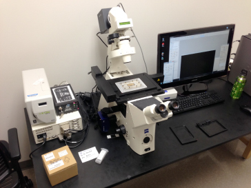
The second major step was the successful design and construction of a new Zeiss microscope, digital camera and anti-vibration table system on which the GeoBioCell is now being operated in the UIUC Institute for Genomic Biology (Fig. 1). This Zeiss Axio Observer inverted microscope is configured for both fluorescence and transmitted light imaging. The fluorescence light path consists of an X-Cite fluorescence lamp coupled to the microscope with a liquid light guide followed by a condenser and filter cubes. The Microscope has three objectives a 10X 0.3NA Neofluar Phase, 20X 0.4NA Plan Neofluar Phase and a 40X 0.6NA Phase LD Plan Neofluar for deep imaging. Images are captured on a 13 magapixel Axiocam HRM black and white camera. For transmitted light imaging, the microscope has a lamp and a 0.55 NA condenser with three phase rings and two DIC settings.
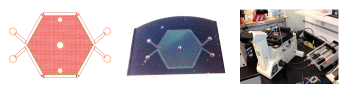
The initial design for the GeoBioCell is based on work done by Zhang et al., 2011 (Fig. 2). The device is designed to mimic naturally occurring microbial niches by capturing features that give rise to rapid emergence of antibiotic resistance in more complex, heterogeneous, environments. The cells are fabricated in silicon wafers. The design is a 1200 hexagonal well structure that allows for concentration gradients to be established with steep and mild gradients across the array. Microbes are inoculated into the wells, and nutrients are delivered to the microbes via diffusion from flow channels around the perimeter of the well structure. Solutes can diffuse between the perimeter flow channels and the well structure, but microbes cannot cross between. This is because the two chambers are connected by 400 nanometer-diameter channels that restrict microbial movement.
The device was fabricated in the Micro and Nanotechnology (MNTL) at UIUC using standard photolithography techniques. Images of the flow cell are shown in the Figure section below. We are currently testing the device using clean water and fluorescein to ensure that we can establish a gradient across the 1200 well array. Once this is established, the GeoBioCell will be inoculated with E. coli and adaptation to an antibiotic concentration gradient will be evaluated.
Publications
-
Dong, Y., Kumar, C. G., Chia, N., Kim, P-J., Miller, P. A., Price, N. D., … Fouke, B. W. (2013). H alomonas sulfidaeris -dominated microbial community inhabits a 1.8 km-deep subsurface Cambrian Sandstone reservoir. Environmental Microbiology, 16(6), 1695–1708. doi:10.1111/1462-2920.12325
-
PROJECT INVESTIGATORS:
-
PROJECT MEMBERS:
Bruce Fouke
Project Investigator
Robert Sanford
Co-Investigator
Charles Werth
Co-Investigator
Reinaldo Alcalde
Collaborator
Laura DeMott
Collaborator
Lang Zhou
Collaborator
-
RELATED OBJECTIVES:
Objective 3.2
Origins and evolution of functional biomolecules
Objective 3.4
Origins of cellularity and protobiological systems
Objective 4.1
Earth's early biosphere.
Objective 4.2
Production of complex life.
Objective 5.1
Environment-dependent, molecular evolution in microorganisms
Objective 5.2
Co-evolution of microbial communities
Objective 5.3
Biochemical adaptation to extreme environments
Objective 6.1
Effects of environmental changes on microbial ecosystems