2014 Annual Science Report
 University of Wisconsin
Reporting | SEP 2013 – DEC 2014
University of Wisconsin
Reporting | SEP 2013 – DEC 2014
Project 4A: New in Situ Techniques (CLSM and Raman) Solve the Problem Presented by the Disaggregation of Acid-Macerated Organic-Walled Microfossils
Project Summary
The search for evidence of past life in rocks to be returned from Mars seems likely to hinge on the use of non-intrusive, non-destructive techniques that can establish the biogenicity of any detected fossil-like objects by analyses both of their cellular morphology and molecular composition. The most promising rock types to preserve such evidence are chemically precipitated sediments such as cherts, gypsums, carbonates and phosphates — examples of all of which on Earth have been shown to be richly fossiliferous. The organic-walled microbes in such rocks are typically not amenable to investigation by the commonly used but rock-destroying technique of acid maceration. This study shows that the combined use of optical microscopy, confocal laser scanning microscopy, and Raman spectroscopy solves this problem, documenting effective means for the investigation of Mars rocks.
Project Progress
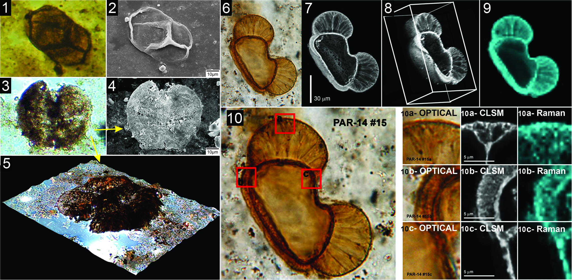
Although the most life-like, “best preserved” fossils in the geological record are those permineralized (“petrified”) in quartz, calcite, apatite or gypsum, the vast majority (>99%) of organic-walled microfossils (e.g., bacteria, fungi, microalgae, acritarchs, spores and pollen) are preserved as compressions, two-dimensional flattened remains compressed between layers of siltstone or shale. Studies of such compression-preserved fossils in situ are far less informative than such studies of three dimensionally preserved permineralized fossils. Nevertheless, compression-preserved microfossils are widespread and especially well known, studied intensively by micropaleontologists worldwide. To identify such specimens and elucidate their morphology, most workers dissolve the fossiliferous rocks in mineral acids (e.g., HF, HCl) and study the macerated acid-resistant organic residues by use of optical (see accompanying figure, parts 1, 2) or scanning electron microscopy (parts 2, 4, 5). Although appropriate for compression-preserved fossils, this technique (as shown by Co-Investigator Ciéber Pereira Caiça, Univ. Sao Paulo and Brazilian Astrobiology Center) does not work well for permineralized fossils, the cell walls of which are mineral-infused, supported by the embedding mineral matrix which as it dissolves during acid maceration commonly results in their disaggregation.
Comparison of results obtained from studies of permineralized ~278 Ma pine pollen in acid-macerates and embedded within the rock matrix illustrates both the problem and its solution. Freed from their embedding matrix, the permineralized acid macerated pollen grains collapse, flatten and provide minimal useful information (accompanying figure, parts 1-5). In contrast, much useful information at high spatial resolution can be obtained in situ by the use of three non-intrusive, non-destructive techniques: optical microscopy (parts 6, 10, 10a-, 10b-, 10c-Optical), confocal laser scanning microscopy, “CLSM” (parts 7, 8, 10a-, 10b-, 10c-CLSM) and Raman spectroscopy (parts 9, 10a-, 10b-, 10c-Raman).
The combined use of these three techniques has numerous advantages:
(1) Unlike the two-dimensional images provided by standard optical photomicrography, structural information provided by CLSM images is three-dimensional and can be rotated and viewed from any desired perspective (compare in the accompanying figure, parts. 6, 7 and 8).
(2) Unlike SEM, which for acid-macerated fossils provides topographic images of the exterior surface of flattened specimens (parts 4 and 5), CLSM images of specimens that transect the thin section surface provide fine-structural detail of the interior structure of cell walls (e.g., parts 10b-CLSM) at a lateral spatial resolution of ~200 nm, 50% higher than standard optical microscopy (compare the optical, CLSM and Raman images in parts 10a, 10b, and 10c).
(3) Because in these specimens CLSM detects laser-induced fluorescence emanating from the polycyclic aromatic hydrocarbons that dominate the carbonaceous kerogen of their cell walls, CLSM serves as a proxy for analyses of chemical composition (compare parts 7 and 9).
(4) Raman spectroscopy provides spectral data that establish the chemical composition of the embedding mineral matrix as well as that of the kerogenous cell walls (blue in parts 9, 10-a, 10b- 10c-Raman).
-
PROJECT INVESTIGATORS:
-
PROJECT MEMBERS:
James Schopf
Project Investigator
Ciéber Caiça
Co-Investigator
Amanda Garcia
Co-Investigator
Anatoliy Kudryavtsev
Co-Investigator
Thomas Fairchild
Collaborator
-
RELATED OBJECTIVES:
Objective 4.1
Earth's early biosphere.
Objective 7.1
Biosignatures to be sought in Solar System materials