2014 Annual Science Report
 University of Wisconsin
Reporting | SEP 2013 – DEC 2014
University of Wisconsin
Reporting | SEP 2013 – DEC 2014
Project 3F -- Apatitic Latest Precambrian and Early Cambrian Fossils Provide Direct Evidence of Concentrations of Environmental Oxygen
Project Summary
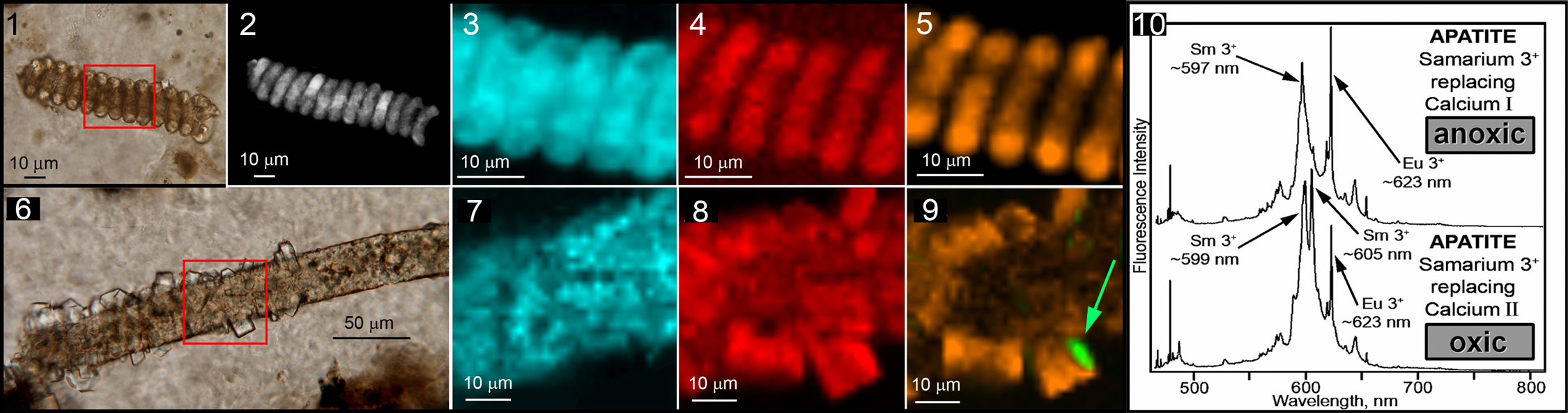
Means are not currently available to asses either quantitatively or semi-quantitatively the concentration of oxygen in Earth’s atmosphere over geological time. Despite this, the environmental availability of O2 has been repeatedly postulated to be a cause of major changes in Earth’s biota, most particularly at the Precambrian-Cambrian boundary-defining “Cambrian Explosion of Life,” a time in Earth history when large deposits of phosphate-rich apatite were deposited in shallow basins worldwide. This study shows that substitution of Sm+3 in the Ca I and Ca II sites of fossil-permineralizing, -infilling, and -encrusting apatite can differentiate between oxic, dysoxic, an anoxic settings of apatite formation. Further studies are to be undertaken to establish such REE-substitution as a quantitative O2 paleobarometer.
Project Progress
Coupled with geologic evidence of the paleoenvironmental setting of the fossil-bearing cherts, the spectroscopic fluorescence data from the Chulaktau fossils (see accompanying figure) are readily explicable: Before microbial decay and disintegration, permineralizing and infilling apatite was emplaced in the low-oxygen (dysoxic) environment of basinal waters at and near the sediment-water interface (resulting in Sm+3-replacement of a mixture of the Ca I and Ca II lattice sites). After burial in unconsolidated anoxic mud, permeating waters carried in phosphate that emplaced fossil-encrusting apatite crystals (and Sm+3-replacement of their Ca I lattice site). At some later time, due to an influx of oxygen-containing percolating waters, the peripheries of some encrusting crystals became oxidized (resulting in replacement at the Ca II lattice site).
For the first time, such data hold promise for deciphering the concentration of dissolved oxygen present in a local apatite-permineralizing, -infilling and -encrusting environment, data of relevance not only to understanding the taphonomy of apatite-associated fossils but of particular importance to understanding the Precambrian-Cambrian boundary-defining “Cambrian Explosion of Life,” an event widely postulated to have coincided with a global increase in environmental oxygen and a time when economically important apatitic phosphate-rich and commonly fossiliferous strata were deposited in shallow basins worldwide.
Initially, our interpretation of variations in apatite fluorescence spectra as being a result of the pattern of lattice site replacement — and, therefore, an indicator of oxic, dysoxic or anoxic conditions — was based on comparison of published spectra with our data from four Neoproterozoic and lowermost Cambrian units (from Alaska, Kazakhstan and southeast Asia). By conducting heating experiments to induce the substitution Sm3+ and other rare-earth elements into the hydroxyapatite lattice under oxygen, inert gas, and vacuum and analyzing the resulting minerals by use of the the same laser line (457.9 nm) used in our earlier analyses we have confirmed our interpretation, showing unambiguously that the fluorescent spectra of samarium-substituted hydroxyapatite can be used to indicate the oxic/dysoxic/anoxic setting in which it formed. Our current goal is to repeat these experiments at 1%, 0.1%, and 0.01% oxygen to provide a quantitative O2 paleobarometer. Results of these studies will be published in an appropriate optical physics journal (e.g., Optical Materials, Journal of Luminescence.)
-
PROJECT INVESTIGATORS:
-
PROJECT MEMBERS:
James Schopf
Project Investigator
Anatoliy Kudryavtsev
Co-Investigator
Vladimir Sergeev
Collaborator
-
RELATED OBJECTIVES:
Objective 4.1
Earth's early biosphere.
Objective 4.2
Production of complex life.
Objective 6.1
Effects of environmental changes on microbial ecosystems
Objective 6.2
Adaptation and evolution of life beyond Earth
Objective 7.2
Biosignatures to be sought in nearby planetary systems