2009 Annual Science Report
 University of Wisconsin
Reporting | JUL 2008 – AUG 2009
University of Wisconsin
Reporting | JUL 2008 – AUG 2009
New Frontiers in Micro-Analysis of Isotopic Compositions of Natural Materials: Development of O, S, Si, and Li Isotopes
Project Summary
The isotope ratios of oxygen and silicon are a sensitive monitor of sedimentary and hydrothermal processes for deposition of banded iron formation. Our focus on banded iron formations reflects the importance these unusual units have in biogeochemical cycling in the early Earth. In particular, we are examining the deposition of microlaminated sections of the Dales Gorge member of the Brockman Iron Formation from the Hamersley basin, which have alternatively been interpreted to represent annual varves in chemical precipitates from seawater, or variations in a sub-surface hydrothermal system. These conflicting models have relevance for interpreting the role of microbial life in precipitation of the Fe-oxides. In addition, δ18O and δ34S values from coexisting minerals can be used to estimate the temperature of the Archean ocean, or of the hydrothermal system. Values of δ7Li and δ18O in zircons allow these tests to be applied to magmas that may have assimilated sedimentary materials, and in the case of pre-4 Ga Jack Hills zircons from SW Australia, provide a record of the earliest Earth before the formation of all known rocks.
Project Progress
Using the University of Wisconsin’s new CAMECA ims-1280 ion microprobe, we have developed the ability to make analyses of: δ18O and δ34S from spots that are 1 to 10 μm in diameter with precision as good as 0.2‰ (2sd, Valley and Kita 2009, Kita et al. 2009, Kozdon et al. 2010); δ30Si in quartz with precision of 0.2‰ (Richter et al. 2009, Knight et al. 2009, Heck et al. 2010); and δ7Li in zircons that contain as little as 1 ppm Li (Ushikubo et al. 2008).
Isotope standards have been developed for O and Si in quartz, S in pyrite, and Li in zircon. We are working to develop standards for O in selected minerals, including magnetite and for S in sphalerite. We discovered that instrument bias varies by up to 5‰ with crystal orientation in the sample mount for δ18O in magnetite (Figure 1, Huberty et al. 2009, 2010, Kita et al. 2009, Valley and Kita 2009, Kozdon et al. 2010). We have found that the magnitude of this effect is significantly less for novel analysis conditions (shallow pits or low accelerating voltage) and we are continuing experiments to optimize analysis protocols. We have also shown that orientation effects do not measurably affect our ability to analyze δ18O in common silicate and carbonate minerals or δ34S in pyrite.
We have investigated and solved several other potentially confounding problems relating to quality of in situ stable isotope analysis. For conditions at WiscSIMS, accuracy and precision are measurably degraded if samples are further than 5mm from a comparative standard. To eliminate such X-Y effects, standards are routinely mounted in the center of every sample. We also find that polishing relief can substantially degrade both precision and accuracy (Figure 2) and that this can be eliminated by careful grinding and polishing procedures. Samples are routinely checked for relief at sub-micron scale using a new white light profilometer. The relief effect is especially significant at grain boundaries. Figure 3 shows an example of optimized data for a well-prepared sample of diopside cast in softer epoxy. The relief is less than 1 μm and analyses on the grain boundary are identical within error of 0.1‰ to analyses from the interior of the grain.
Detailed petrography by optics and SEM-BSE is underway on BIFs samples from Dales Gorge, Kuruman (South Africa), Isua, and Biwabik/Gunflint; work is also planned for the 3.5 Ga Apex and Strelley Pool cherts. These samples will be investigated by in situ stable isotope analysis during the coming year.
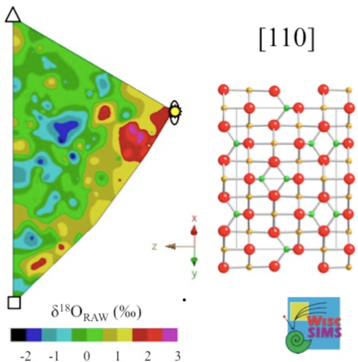
Figure 1. Raw values of oxygen isotope ratio measured by SIMS in magnetite vary by 5‰ for different crystallographic orientations within the sample mount. This orientation effect can be minimized with special tuning of the ion microprobe (Huberty et al. 2009, 2010). Orientation effects are not detectable for common silicate and carbonate minerals (Valley and Kita 2009).
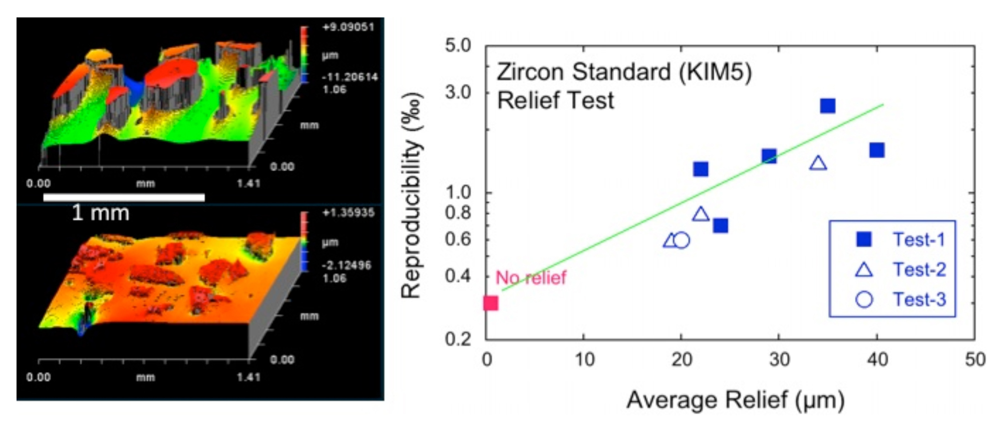
Figure 2. Tests of the effect of polishing relief on reproducibility of oxygen isotope ratios measured from 10 μm spots by ion microprobe in zircon grains cast in epoxy. Grains with less than 2 μm of relief are precise to ±0.3‰ (2SD), while grains with 30-40 μm of relief are shifted in value by 3-5‰ and 10 times less precise, ±3‰ (Kita et al. 2009).
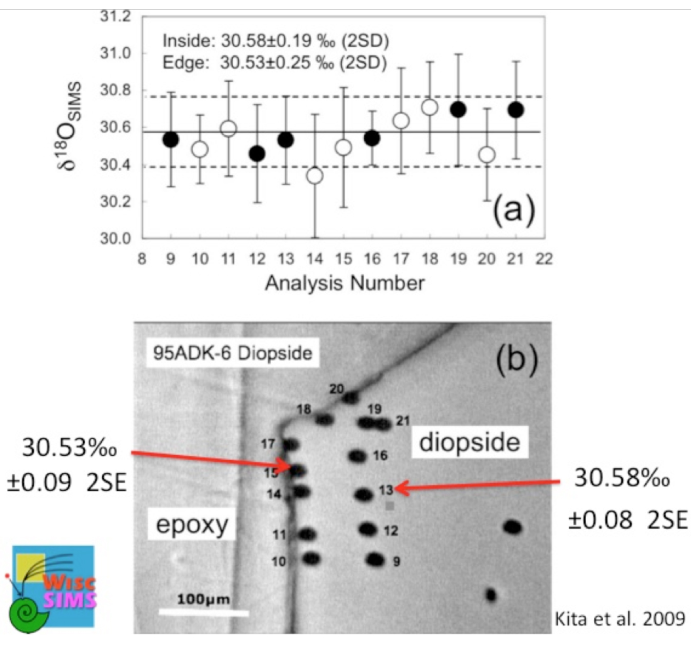
Figure 3. Highly precise and accurate analysis of oxygen isotope ratios is achieved even for samples of different hardness if polished carefully. Analyses of diopside standard cast in epoxy are identical within 0.1‰ for grain boundaries against epoxy and interiors (Kita et al. 2009).
Publications
-
Blank, J. G., Green, S. J., Blake, D., Valley, J. W., Kita, N. T., Treiman, A., & Dobson, P. F. (2009). An alkaline spring system within the Del Puerto Ophiolite (California, USA): A Mars analog site. Planetary and Space Science, 57(5-6), 533–540. doi:10.1016/j.pss.2008.11.018
-
Dattagupta, S., Schaperdoth, I., Montanari, A., Mariani, S., Kita, N., Valley, J. W., & MacAlady, J. L. (2009). A novel symbiosis between chemoautotrophic bacteria and a freshwater cave amphipod. ISME J, 3(8), 935–943. doi:10.1038/ismej.2009.34
-
Huberty, J. M., Kita, N. T., Kozdon, R., Heck, P. R., Fournelle, J. H., Spicuzza, M. J., … Valley, J. W. (2010). Crystal orientation effects in δ18O for magnetite and hematite by SIMS. Chemical Geology, 276(3-4), 269–283. doi:10.1016/j.chemgeo.2010.06.012
-
Kita, N. T., Ushikubo, T., Fu, B., & Valley, J. W. (2009). High precision SIMS oxygen isotope analysis and the effect of sample topography. Chemical Geology, 264(1-4), 43–57. doi:10.1016/j.chemgeo.2009.02.012
-
Knight, K. B., Kita, N. T., Mendybaev, R. A., Richter, F. M., Davis, A. M., & Valley, J. W. (2009). Silicon isotopic fractionation of CAI-like vacuum evaporation residues. Geochimica et Cosmochimica Acta, 73(20), 6390–6401. doi:10.1016/j.gca.2009.07.008
-
Kozdon, R., Kita, N. T., Huberty, J. M., Fournelle, J. H., Johnson, C. A., & Valley, J. W. (2010). In situ sulfur isotope analysis of sulfide minerals by SIMS: Precision and accuracy, with application to thermometry of ∼3.5Ga Pilbara cherts. Chemical Geology, 275(3-4), 243–253. doi:10.1016/j.chemgeo.2010.05.015
-
Richter, F. M., Watson, E. B., Mendybaev, R., Dauphas, N., Georg, B., Watkins, J., & Valley, J. (2009). Isotopic fractionation of the major elements of molten basalt by chemical and thermal diffusion. Geochimica et Cosmochimica Acta, 73(14), 4250–4263. doi:10.1016/j.gca.2009.04.011
- Huberty, J.M., Kita, N.T., Heck, P.R., Kozdon, R., Fournelle, J.H., Xu, H. & Valley, J.W. (2009). Crystal orientation effects on bias of δ18O in magnetite by SIMS. Goldschmidt Conference. Geochim. Cosmochim. Acta, 73(13).
- Kita, N.T., Huberty, J.M., Kozdon, R., Beard, B.L. & Valley, J.W. (2009, In Review). High precision SIMS oxygen, sulfur and iron stable isotope analyses of geological materials: Accuracy, surface topography and crystal orientation. SIMS XVII Proceedings (Special Issue, Surface and Interface Analysis).
- Ushikubo, T., Kita, N.T., Cavosie, A.J., Wilde, S.A., Rudnick, R.L. & Valley, J.W. (2008). Lithium in Jack Hills zircons: Evidence for extensive weathering of Earth’s earliest crust. Earth and Planetary Science Letters, 272: 666-676.
- Valley, J.W. & Kita, N.T. (2009). In situ Oxygen Isotope Geochemistry by Ion Microprobe. In: Fayek, M. (Eds.). AC Short Course: Secondary Ion Mass Spectrometry in the Earth Sciences. Vol. 41.
-
PROJECT INVESTIGATORS:
-
PROJECT MEMBERS:
Huifang Xu
Project Investigator
Philipp Heck
Postdoc
Reinhard Kozdon
Postdoc
Takayuki Ushikubo
Postdoc
John Fournelle
Research Staff
Brian Hess
Research Staff
Jim Kern
Research Staff
Mike Spicuzza
Research Staff
Jason Huberty
Graduate Student
-
RELATED OBJECTIVES:
Objective 4.1
Earth's early biosphere.
Objective 7.1
Biosignatures to be sought in Solar System materials

