2009 Annual Science Report
 University of Wisconsin
Reporting | JUL 2008 – AUG 2009
University of Wisconsin
Reporting | JUL 2008 – AUG 2009
Development of Laser Ablation-Electron Impact Ionization-Miniature Mass Spectrometer (LA-EI-MMS) for In-Situ Chemical and Isotopic Measurements of Martian Rocks
Project Summary
Our goal is to develop instrumentation capable of being deployed on a lander style space craft that can measure the chemical and isotopic composition of Martian samples. The instrumentation is based on the technologies of (i) laser ablation (LA) sampling of minerals, (ii) electron-impact ionization (EI) of ablated neutrals, and (iii) mass spectral measurement using JPL-developed miniature mass spectrometer (MMS) of focal plane geometry with modified-CCD array detector.
Project Progress
The instrument performs the mass spectral analysis of the laser ablated vapors from solids using a miniature mass spectrometer (MMS) of focal plane geometry with modified CCD based array detector for the direct and simultaneous measurement of different mass ions.
Figure 1 shows the technical approach schematically and Figure 2 shows a photograph of the working mass spectrometer with modified CCD detector array. The working mass spectrometer is approximately 1.5kg in mass which includes housing power supply and pump, it uses 3-5 W of power in operation, has a mass resolving power of ~315 and can simultaneously analyzed from mass 2 to 250 with a dynamic range of 104 and an estimated minimum detection limit of 50 ions presently.

Figure.1: The vapor of the neutral species ablated by the laser pulse is led into the electron-impact ion source. The ions thus generated are analyzed by the miniature mass spectrometer with modified CCD ion detector array.

Figure 2: Photograph of the working miniature mass spectrometer with modified CCD ion detector array.
We have made mass spectral measurements of different rock samples by laser ablation-mass spectrometry. The samples were places in the mass spectrometer chamber. The output of Nd-YAG laser (λ =1.06 µm) was focused onto the rock. The ablated vapor was inlet into the electron impact ionization source and neutral vapor species after ionization were mass analyzed by the MMS and CCD direct ion detector array. The different mass ions are spatially separated along the focal plane of the magnetic sector. The intensities of the ions are simultaneously measured by the array detector. CCD detector is an integrating detector and integrates the ions impinging on them for the specified period. Results of two tests are reported below that show the miniature mass spectrometers ability to determine the chemical composition of a complex rock (a dacitic volcanic glass) and the C isotope composition of calcite (CaCO3)
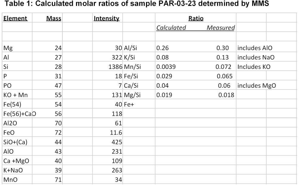
Figure 3 shows the mass spectral analysis of a dacitic volcanic glass sample measured using the miniature mass spectrometer by electron impact ionization of laser ablated neutral atoms. The mass spectrum is complicated and includes elemental and oxide peaks as well as some peaks at low mass that are unknown (e.g., unidentified peaks at mass 18 and 26). However by normalizing the peaks to Si (mass 28) ion intensity and combining the oxide and elemental peaks the measured molar ratios relative to calculated ratios determined by x-ray fluorescence is good as listed in table 1.
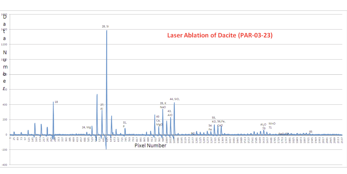
Figure 3: Plot of relative ion intensity versus mass showing the mass spectrum of laser ablation of a volcanic rock

Figure 4: Plot of relative ion intensity versus mass showing the two major CO peaks from laser ablation of calcite and the inferred C isotope composition.
The ability of the miniature mass spectrometer to determine the isotopic composition of an element is shown by the C isotope analysis of calcite. The mass spectrum acquired by laser ablation is shown in figure 4 in which the 13C/13C ratio was determined from the ratio of mass 45 to mass 44 in which it is assumed that only 16O is present in these two CO2 peaks.
One of the most significant impediments to this work has been cause by the laser pulse producing changes in the voltages of the source ion optics. These voltage fluctuations are thought to arise from production of high potential ions produced by laser sputtering. It is these ion optic voltage fluctuations that are the likely cause of some of the error in the ion intensity measurements.
Future Plan:
Much of the calibration work that is needed will be conducted Nov/Dec of 2009 at which time Jim Ludois a UW astrobiology graduate student will spend 3 weeks at JPL making chemical analyses of a variety of minerals to evaluate the complex elemental and oxide spectrum produced by the laser ablation system. Additionally we will begin to evaluate the capabilities of the mass spectrometer system for the isotopic analysis of major elements including C, O, and Fe.
Other items devoted to the development of this instrumentation include:
● Overcome the laser pulse effect on the ion source and detector by implementing ion repeller screens and detector shielding.
● Modify the chamber to allow for easier alignment of the laser, target and the mass spectrometer and synchronize laser pulsing with signal integration time to facilitate easier data acquisition
● Improve detector performance for sensitivity to allow isotopic analysis of low abundance ions, and evaluate inter-channel gains in the modified CCD
-
PROJECT INVESTIGATORS:
-
PROJECT MEMBERS:
Clark Johnson
Collaborator
Jim Ludois
Graduate Student
-
RELATED OBJECTIVES:
Objective 2.1
Mars exploration.
Objective 7.2
Biosignatures to be sought in nearby planetary systems
