2010 Annual Science Report
 University of Wisconsin
Reporting | SEP 2009 – AUG 2010
University of Wisconsin
Reporting | SEP 2009 – AUG 2010
Project 1C: Extra-Cellular Polymeric Substances as Armor Against Cell Membrane Rupture on Mineral Surfaces
Project Summary
Our interdisciplinary project examined the hypotheses that bacterial cell membranes are ruptured in contact with specific mineral surfaces, and that biofilm-forming extra-cellular polymeric substances (EPS) may have evolved to shield against membrane rupture (cell lysis). Furthermore, we proposed that mineral reactivity towards membranolysis should depend on its surface properties such as charge, reactive area, or free radicals generated by radiation and impacts on early Earth, Mars, and other worlds. The effect of EPS on preservation in the rock record will also be examined. By understanding the mechanisms for membranolysis, especially under the extreme conditions of high radiation and heavy impacts during early planetary history, the project addresses the NASA Astrobiology Institute’s (NAI) Roadmap goals of understanding the origins of cellularity, the evolution of mechanisms for survival at environmental limits, and preservation of biosignatures, and NASA’s Strategic Goal of advancing scientific knowledge of the origin and evolution of the Earth’s biosphere and the potential for life elsewhere.
Project Progress
We used whole bacterial cells to address the question of whether EPS provides armor against cell lysis. A newly developed EPS-lacking mutant strain of Pseudomonas aeruginosa (Figure 1), and the wild-type, which is a robust EPS producer (Figure 1), were used to conduct cell viability studies in suspensions of oxide particles with cells. Wild type cell viability was greater than that of the mutant type. Furthermore, viability was oxide-dependent, increasing as γ-Al2O3 > amorphous SiO2 (Figure 2). Thus, our results confirm our hypothesis that EPS acts as a shield against the toxicity of certain minerals. We also examined the cell + oxide particle suspensions using High Resolution Transmission Electron Microscopy (HRTEM) to visualize the location of the particles with respect to the cell surface. The amorphous silica particles were located in the extra-cellular region of both wild type cells that possess EPS as well as washed mutant type cells that lack EPS, suggesting that the similar negative charge on both SiO2 particles and the cell surfaces resulted in electrostatic repulsion and prevented cell-particle contact. In contrast, γ-Al2O3 particles of size < 80 nm were seen inside the washed mutant type cells, but were seen only in the extracellular space for the wild type cells. These results indicated that the positively-charged γ-Al2O3 particles were electrostatically attracted to the oppositely-charged cell surface. The particles could not penetrate in the case of the wild type due to the presence of the EPS, whereas the smaller particles < 80 nm could enter the mutant type cells that lack EPS. We are also currently investigating the effect of reactive oxygen species at TiO2 surfaces on cell viability. Our results support our hypotheses that EPS acts an armor against mineral toxicity, and that not all minerals are toxic or benign, but that toxicity depends on mineral surface properties such as surface charge, surface free radicals, etc.
We are currently planning to conduct silicification experiments for both wild and mutant type cells in order to examine differences in morphology for preservation differences.
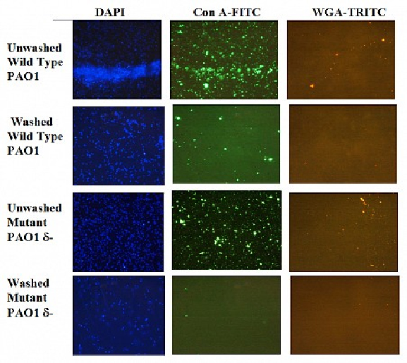
Figure 1. Visualization of Extracellular Polymeric Substances (EPS) Using Fluorescent Stains. Lectin stains for identifying EPS (1000x magnification) of washed and unwashed wild type and mutant type cells of Pseudmonas aeruginosa. Washing removes traces of EPS associated with the cells. DAPI (blue) stains for DNA indicating presence off cells (left); green con A-FTIC (center) and red WGA-TRITC (right) stain for specific EPS (Xu et al., in prep.).

Figure 2. Viability of cells in the presence of amorphous SiO2 (a) and gamma-Al2O3 (b) particle suspensions normalized to oxide-free controls, for cells grown in Luria-Betani culture medium and suspended in 0.5% saline solution. Note that viability of wild type cells (WT) > washed wild-type (WWT) > washed mutant type (WMT), for both oxides, and the differences are more marked for gamma-Al2O3. Thus, absence of EPS decreases viability. Also note that viability is strongly dependent on oxide, with gamma-Al2O3 being much more toxic than amorphous SiO2 (Xu et al., in prep.)

Figure 3. HRTEM images of WT (a) and wMT (b) cells in amorphous SiO2 suspensions, and WT© and wMT (d) cells gamma-Al2O3 suspenions. Note that the SiO2 particles do not enter either type of cell, whereas gamma-Al2O3 particles < ~ 80 nm penetrate into the wMT cells (d) but not WT cells©. The cell surface is negatively charged so the negatively-charged SiO2 particles are electrostatically repelled and do not enter either WT or wMT cell. In contrast, the positively-charged gamma-Al2O3 particles are attracted to the oppositely-charged cell surface, and the smaller particles can penetrate into the intracellular region and, presumably, affect cell viability by interfering with normal cell function. Scale bar = 200 nm.
Publications
-
Xu, J., Stevens, M. J., Oleson, T. A., Last, J. A., & Sahai, N. (2009). Role of Oxide Surface Chemistry and Phospholipid Phase on Adsorption and Self-Assembly: Isotherms and Atomic Force Microscopy. The Journal of Physical Chemistry C, 113(6), 2187–2196. doi:10.1021/jp807680d
- Xu, J., Sahai, N. & Schoonen, M.A. (2010, in Preparation). Exploring the mechanisms for highly reactive oxygen species (HROS) formation at oxide particle surfaces in aqueous suspensions: Implications for the origin and evolution of life at mineral surfaces. Astrobiology.
- Xu, J., Sahai, N., Hickey, W.J. & Capbell, J. (2010, in Preparation). Extra-cellular polymeric substances as armor against cell membrane rupture on mineral surfaces. Nature.
-
PROJECT INVESTIGATORS:
-
PROJECT MEMBERS:
William Hickey
Co-Investigator
Martin Schoonen
Co-Investigator
Jie Xu
Doctoral Student
-
RELATED OBJECTIVES:
Objective 3.4
Origins of cellularity and protobiological systems
Objective 5.1
Environment-dependent, molecular evolution in microorganisms
Objective 7.1
Biosignatures to be sought in Solar System materials
