2010 Annual Science Report
 Georgia Institute of Technology
Reporting | SEP 2009 – AUG 2010
Georgia Institute of Technology
Reporting | SEP 2009 – AUG 2010
Experimental Model System - an Ancestral Magnesium-RNA-Peptide Complex
Project Summary
We are developing small model systems in which the interactions of rPeptides, Mg2+ ions and rRNA can be studied by NMR, X-ray diffraction, calorimetry, molecular dynamics simulations, and other ‘high resolution’ biophysical techniques. Within the large subunit of the extant ribosome, one observes an autonomous rRNA:Mg2+-mc complex in Domain III, which appears to fold independent of the rest of the LSU. Ribosomal protein L23 associates closely with this rRNA: Mg2+-mc complex in both bacteria and archaea, suggesting the possibility of distinct evolutionary origin. We will define the smallest Domain III rRNA and associated peptide segments sufficient for assembly of this complex, and will characterize their assembly and interactions with a-PTC and 23S lacking Domain III by a variety of experimental and computational methods.
Project Progress
We are developing systems to deconstruct the extant LSU so that we can study the molecular interactions within it. Noller, 1 Nierhaus, 2 and others showed that extant LSU ribosomal assembly requires only ribosomal proteins L2, L3 and L4, the 23S rRNA, along with inorganic ions such as Mg2+, K+, and Na+. Our model of the a-PTC contains a-rRNA and three a-rPeptides (a-PL2, a-PL3, and a-PL4), with sequences corresponding to the L2, L3, and L4 tails that drill deeply into the LSU. The a-PTC contains six highly coordinated Mg2+ ions, which are in the form of three “magnesium microclusters” (Mg2+-μc’s). The sequences and molecular interactions of these ancestral components are conserved over vast evolutionary timescales, predating the last common universal ancestor of life. 3, 4 Our structural analysis and published biochemical results5 indicated that one of the Mg2+-μc’s and coordinated a-rRNA is self-assembling.
Exclusive of our model a-PTC, a Mg2+-μc in Domain III (Figure 1) is unique in its autonomy within LSU. Consistent with the Bokov assembly model 6, the Domain III rRNA:Mg2+-μc complex appears to fold independent of other LSU rRNA, but integrates closely with rPeptide PL39 in the H. marismortui crystal structure and rPeptide PL23 in the T. thermophilus and H. marismortui structures. Most of the PL23 structure in complex with the T.thermophilus Domain III aligns closely with PL23 in H. marismortui when the rRNA and Mg2+-μc’s are superimposed. As coordination of the Mg2+-μc in Domain III is contained entirely within this domain, an independent fold and distinct evolutionary origin seems possible, if not likely. However, the “tail” region of PL23 in T.thermophilus extends into and creates part of the PTC exit tunnel, whereas the “tail” region of H. marismortui’s PL23 is much shorter. Unlike T.thermophilus, a second protein (rPeptide PL39) associates with Domain III of H. marismortui and extends into the PTC exit tunnel, possibly replacing PL23. Our team’s sequence mining and consensus analyses on PL23 and PL39 suggest that the “tail” region of PL23 observed to create part of the PTC exit tunnel in T.thermophilus appears characteristic of all bacteria and absent in archaea and eukaryotes. Additionally, PL39 is fairly conserved among eukaryotes and archaea, and entirely absent in bacteria.
Though independent of the model a-PTC, Domain III in the large subunit of the extant ribosome is a promising experimental small model system. One of two initial model systems of the Domain III rRNA has been synthesized (in DNA form) and replicated in preparation for transcription. Our goals for this model system are to characterize its ability to fold and assemble as a small model system, which will facilitate an integrated experimental and computational investigation of its interactions and assembly, and later to evaluate its interactions with the model a-PTC and 23S lacking Domain III.
1. Khaitovich, P.; Mankin, A. S.; Green, R.; Lancaster, L.; Noller, H. F., Characterization of functionally active subribosomal particles from Thermus aquaticus. PNAS 1999, 96 (1), 85-90.
2. Schulze, H.; Nierhaus, K. H., Minimal set of ribosomal components for reconstitution of the peptidyltransferase activity. EMBO J. 1982, 1, 609-613.
3. Hsiao, C.; Mohan, S.; Kalahar, B. K.; Williams, L. D., Peeling the onion: Ribosomes are ancient molecular fossils. Molecular Biology and Evolution 2009, 26 (11), 2415-2425.
4. Hsiao, C.; Tannenbaum, M.; VanDeusen, H.; Hershkovitz, E.; Perng, G.; Tannenbaum, A.; Williams, L. D., Complexes of Nucleic Acids with Group I and II Cations. In Nucleic Acid Metal Ion Interactions, Hud, N., Ed. The Royal Society of Chemistry: London, 2008; pp 1-35.
5. Kitahara, K.; Kajiura, A.; Sato, N. S.; Suzuki, T., Functional genetic selection of helix 66 in Escherichia coli 23S rRNA identified the eukaryotic-binding sequence for ribosomal protein L2. Nucleic Acids Research 2007, 35 (12), 4018-4029.
6. Bokov, K.; Steinberg, S. V., A hierarchical model for evolution of 23S ribosomal RNA. Nature 2009, 457 (7232), 977-980.
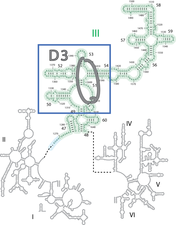
Figure 1. Domain III (outset and enlarged) from the 23S secondary structure of T. thermophilus. Intra-domain rRNA:Mg2+-µc coordination is shown in grey.
Publications
-
Bokov, K., & Steinberg, S. V. (2009). A hierarchical model for evolution of 23S ribosomal RNA. Nature, 457(7232), 977–980. doi:10.1038/nature07749
-
Hsiao, C., Mohan, S., Kalahar, B. K., & Williams, L. D. (2009). Peeling the Onion: Ribosomes Are Ancient Molecular Fossils. Molecular Biology and Evolution, 26(11), 2415–2425. doi:10.1093/molbev/msp163
-
Kitahara, K., Kajiura, A., Sato, N. S., & Suzuki, T. (2007). Functional genetic selection of Helix 66 in Escherichia coli 23S rRNA identified the eukaryotic-binding sequence for ribosomal protein L2. Nucleic Acids Research, 35(12), 4018–4029. doi:10.1093/nar/gkm356
- Hsiao, C., Tannenbaum, M., VanDeusen, H., Hershkovitz, E., Perng, G., Tannenbaum, A. & Williams, L.D. (2008). Complexes of Nucleic Acids with Group I and II Cations. In: Hud, N. (Eds.). Nucleic Acid Metal Ion Interactions. London: The Royal Society of Chemistry.
- Khaitovich, P., Mankin, A.S., Green, R., Lancaster, L. & Noller, H.F. (1999). Characterization of functionally active subribosomal particles from Thermus aquaticus. PNAS, 96(1): 85-90.
- Schulze, H. & Nierhaus, K.H. (1982). Minimal set of ribosomal components for reconstitution of the peptidyltransferase activity. EMBO J, 1: 609-613.
-
PROJECT INVESTIGATORS:
-
PROJECT MEMBERS:
Chiaolong Hsiao
Postdoc
Chad Bernier
Graduate Student
-
RELATED OBJECTIVES:
Objective 3.2
Origins and evolution of functional biomolecules
