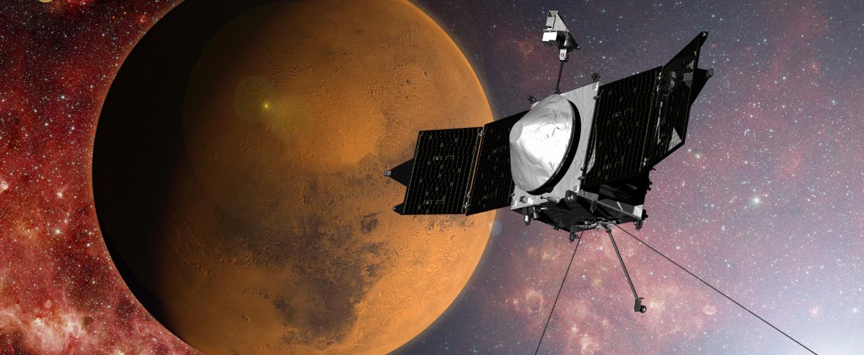
NASA's MAVEN mission carries the Neutral Gas and Ion Mass Spectrometer (NGIMS). Mass spectrometers have numerous applications in astrobiology research, and the additional technique of mass spectrometry Imaging (MSI) could also have an important role in future studies.Image Credit: NASA
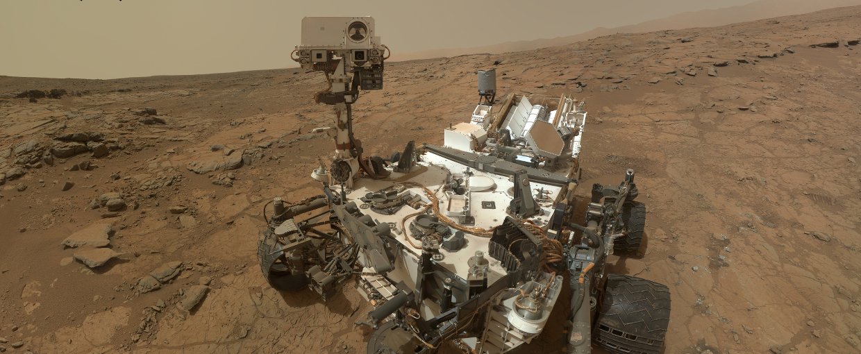
The Sample Analysis at Mars (SAM) instrument on NASA's Curiosity contains a mass spectrometer. Mass spectrometers have numerous applications in astrobiology research, and the additional technique of mass spectrometry Imaging (MSI) could also have an important role in future studies.Image Credit: NASA/JPL-Caltech/MSSS
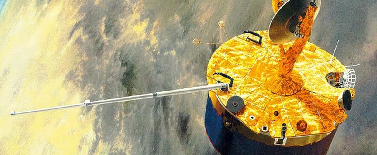
The Pioneer Venus Orbiter Neutral Mass Spectrometer (ONMS) was designed to measure density variations of the major neutral constituents in Venus' upper atmosphere. Mass spectrometers have numerous applications in astrobiology research, and the additional technique of mass spectrometry Imaging (MSI) could also have an important role in future studies.Image Credit: NASA
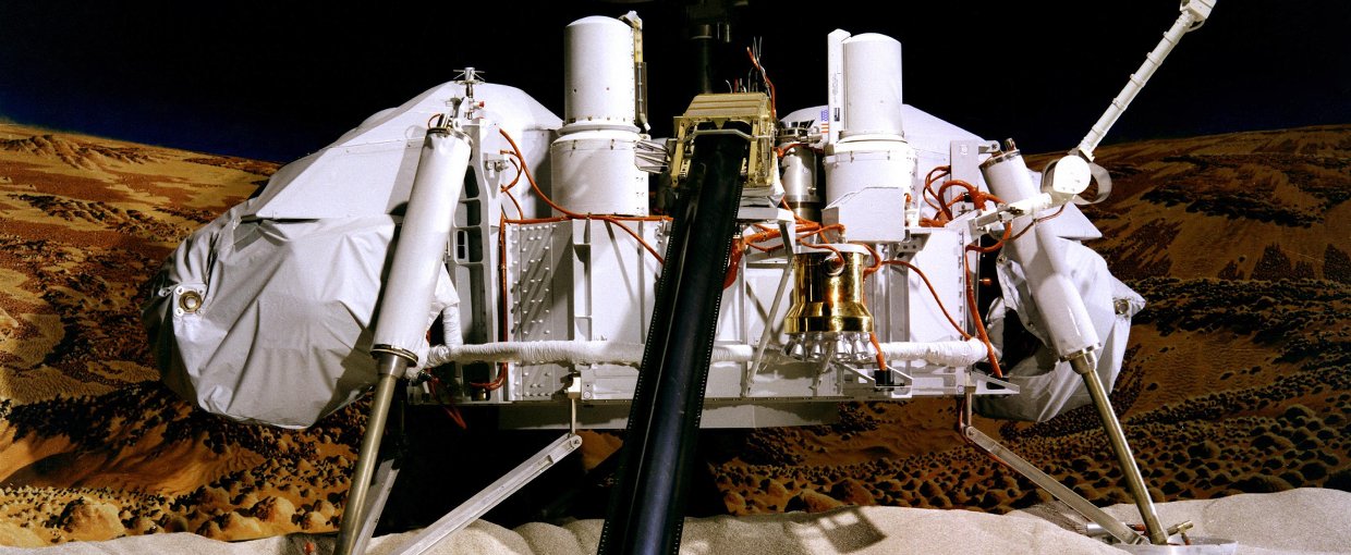
In the 1970s, the Viking 1 and Viking 2 landers each carried a gas chromatograph—mass spectrometer instrument to Mars. Mass spectrometers have numerous applications in astrobiology research, and the additional technique of mass spectrometry Imaging (MSI) could also have an important role in future studies.Image Credit: NASA/JPL-Caltech/University of Arizona
Mass spectrometry is an analytical technique used in countless scientific applications, including space missions ranging from planetary rovers like Curiosity to orbital spacecraft like LADEE and MAVEN. Mass spectrometry works by ionizing chemicals in a sample and sorting the resulting ions based on their mass-to-charge ratio.
An additional technique, known as mass spectrometry imaging (MSI), provides visual information about how chemical compositions are distributed spatially. MSI is a relatively new field and, in recent years, its application has been expanding rapidly. Uses for MSI potentially include studies relevant to astrobiology, such as the visualization of compounds useful as biomarkers.
A recent review discusses the wide range of MSI tools now available, both custom-built and from commercial vendors. The authors focus on well-established, as well as new or emerging, MSI techniques that could prove useful in biomaterial analysis. According to the publication, the aim is to:
“provide a concise resource for those interested in utilizing MSI for applications such as biomimetic materials, biological/synthetic material interfaces, polymer formulation and bulk property characterization, as well as the spatial and chemical distributions of nanoparticles, or any other molecular imaging application requiring broad chemical speciation.”
The paper, “Visualizing molecular distributions for biomaterials applications with mass spectrometry imaging: a review,” was published in the Journal of Materials Chemistry B. The work was performed by the NSF/NASA Center for Chemical Evolution (CCE) at the Georgia Institute of Technology in Atlanta, Georgia. The CCE is a collaborative program supported by the National Science Foundation (NSF) and the NASA Astrobiology Program.