2011 Annual Science Report
 University of Wisconsin
Reporting | SEP 2010 – AUG 2011
Project 7C: Improving Accuracy of in Situ Stable Isotope Analysis by SIMS
Project Summary
Isotopic analysis of biologically important elements in petrographic context is a fundamental tool for astrobiology. Secondary ion mass spectrometry (SIMS) is a powerful tool used to understand biogeochemical processes at the spatial scale of the individual microorganisms that drive them. The benefits of the extremely high spatial resolution offered by SIMS do not come without costs, however. Unconstrained physical and chemical variables in the sample of interest can introduce biases leading to inaccurate measurements. Understanding and constraining these barriers to accuracy as we move towards ever finer spatial resolution and analytical precision is a primary focus at WiscSIMS.
Project Progress
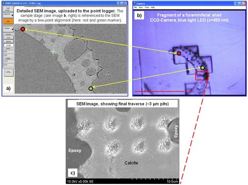
The ability to make small to ultra-small spot (<3 μm) analyses of oxygen and sulfur isotope ratios with a typical spot-to-spot precision of 0.3‰ (2 SD) for 10 µm spot sizes and ~0.7‰ for 3 µm beam spots has been developed and is now routinely performed at WiscSIMS, the University of Wisconsin’s CAMECA ims-1280 ion microprobe laboratory (Kita et al. 2011, Ushikubo, 2010; Williford et al. 2011). Smaller spots are possible at a small trade-off in precision (Page et al. 2007, Nakamura et al 2008, Kozdon et al. 2009, 2010, Kita et al. 2010, Noguchi et al. 2011, Williford et al. 2011). In order to take full advantage of this high spatial resolution, the analysis pit has to be placed as accurately as possible on the sample, however the white light viewing system as delivered by CAMECA had a spatial resolution of ~4 μm and while larger spot analyses can be viewed before, during, and after an analysis, smaller pits were invisible to the analyst and could only be seen after analysis by SEM. We have implemented several modifications to improve and simplify sample navigation and aiming: (1) A point logger software (Figure 1) allows the use of detailed scanning electron microscope (SEM) images or high resolution images acquired by other techniques as reference for precise aiming. (2) The resolution of the optical system for live (optical) imaging was significantly improved by changing the original light source from white light to either a blue or near-ultraviolet LED (λ = 368 nm). Implementation of the UV illumination required that several optical elements were replaced for an enhanced transmission of light in the near-UV range, and a UV-sensitive CCD camera with quadrupled pixels was installed. Together, these modifications assist the operator during aiming of the analysis spot and therefore reduce the idle time between to measurements. (3) Vibration of the sample stage was minimized by retrofitting vibration dampers to turbo pumps (Figure 2). (4) We have installed optical encoders to the stage positioning with sub-micron accuracy of X-Y position.
Our continued efforts to develop procedures for improved accuracy and smaller spots for stable isotope analysis is facilitating studies such as sulfur isotope analysis with pits 3×2 μm in size in finely zoned samples with a precision in δ34S of ±0.6‰ (Williford et al., 2011).
While evaluating new standards, the dependence of measured isotope ratios of oxygen and iron on the crystal orientation of magnetite, oxygen in hematite, and sulfur in sphalerite and galena was discovered and reported in three papers (Huberty et al., 2010; Kita et al., 2011; Kozdon et al., 2010). Measured isotope ratios by SIMS were correlated with crystal orientations of individual grains as determined by electron backscatter diffraction (EBSD).
We developed new analytical protocols to improve the accuracy of SIMS analysis of δ18O in magnetite and hematite and δ34S in sphalerite and galena by reducing both the total impact energy and the incident beam angle of the primary ions. The precision in δ34S from grain-to-grain of sphalerite, which shows an orientation effect on analytical bias, improves from ±1.7‰ (2SD) at routine SIMS analytical conditions to ±0.6‰ by reducing the total impact energy of the primary ions from 20 to 13 keV (Kozdon et al., 2010). For δ18O in magnetite and hematite, precision in δ18O from grain-to-grain improves from ±3‰ (2SD) at routine analytical conditions to ±0.8‰ at 13 keV (Huberty et al., 2010). Thus the affect of crystal orientation on measured isotope ratio can be minimized. While the effect of crystal orientation can be significant for a few minerals, it has been confirmed that orientation effects are not detectable at a precision of ±0.3 ‰ on a wide range of over 50 silicate, carbonate, and sulfide minerals.
Another key development at WiscSIMS is in situ carbon isotope analysis of ancient sedimentary organic matter. Sedimentary organic matter is highly variable in composition and mode of preservation relative to crystalline mineral phases (e.g. pyrite, zircon), and this presents certain analytical challenges. Compositional variability related to organic chemistry or variable inclusion of mineral phases can introduce matrix effects, and the continuum of sizes and concentrations at which sedimentary organic matter is preserved leads to variable ion count rates relative to working standards. A correlation between H/C and instrumental bias for SIMS carbon isotope analysis of organic matter has been reported (Sangély et al., 2005), and we have confirmed and expanded on these results with our own experiments. That study used Fourier transform infrared spectroscopy on each analytical pit to estimate H/C. We have developed a method to measure H/C and δ13C simultaneously from the same pit, which minimizes potential interferences and maximizes efficiency. Using a suite of 12 internal laboratory standard organic materials with H/C (atomic) between 0.1 and 1.7, we find a strong linear correlation over a range of ~4‰. A subset of these standards is analyzed at the beginning of each session in order to calibrate the relationship and correct for the H/C effect in samples of unknown composition. We have achieved an average reproducibility of 0.4‰ (2 SD) using a 6 μm beam diameter with two faraday cup (FC) detectors, and 0.8‰ (2 SD) using a 1 μm beam diameter with a FC for 12C and an electron multiplier (EM) for 13C.
We have also developed a method for measuring in situ δ13C of very low TOC materials including microbial fossils preserved in chert. To achieve ion count rates sufficiently large to ensure accurate analyses of these materials, a larger, higher intensity beam must be used along with FC/EM detectors, and a standard with similar chemical composition to the samples is also critical. When using an EM detector and a high intensity beam, it is impossible to analyze the suite of high TOC standards discussed above (due to EM saturation). Using a carbonaceous chert from the ~3.4 Ga Fig Tree Group of South Africa (House et al., 2000) and a 10–15 mm beam diameter, we achieved an average reproducibility of 1.9‰ (2 SD). Average internal precision for these analyses is 1.3‰ (2 SD), and it is possible that our reproducibility is limited by isotopic heterogeneity in the standard. We are currently developing a suite of carbonaceous chert standards with a range of H/C in order to evaluate this effect using the high intensity beam.
Fig. 1: a-c) Images demonstrating the capabilities of the point logger software for improved sample navigation and aiming. a) Detailed scanning electron microscope (SEM) image of a foraminiferal chamber wall cross section. b) Same sample as shown in (a), imaged by the built-in CCD camera of the SIMS optical system and as seen by the operator using the new blue-light optical system. Depending on the sample type and surface texture, accurate aiming based on this live image may be difficult. With the point logger software, the sample stage can be referenced to the SEM image using a two-point alignment (red and green markers indicate reference points). This allows the selection of analysis-spot locations using features visible on the SEM image instead of relying on the lower resolution live optical image. c) Traverse for δ18O crosscutting a foraminiferal chamber wall featuring pit-placement within 1 to 2 µm accuracy.
References
Dattagupta S, Schaperdoth I, Montanari A, Mariani S, Kita N, Valley JW, and. Macalady JL (2009) A Recently Evolved Symbiosis Between Chemoautotrophic Bacteria and a Cave-dwelling Amphipod, Int. Soc. Microbial Ecology J., 1-9.
House CH, Schopf JW, McKeegan KD, Coath CD, Harrison TM, Stetter KO (2000) Carbon isotopic composition of individual Precambrian microfossils. Geology 28:707-710.
Huberty, J.M., Kita, N.T., Kozdon, R., Heck, P.R., Fournelle, J.H., Spicuzza, M.J., Xu, H., Valley, J.W., 2010. Crystal orientation effects in δ18O for magnetite and hematite by SIMS. Chem. Geol., 276(3-4): 269-283.
Kita NT, Nagahara H, Tachibana S, Tomomura S, Spicuzza MJ, Fournelle JH and Valley JW (2010) High precision SIMS oxygen three isotope study of chondrules in LL3 chondrites: Role of ambient gas during chondrule formation. Geochim. Cosmochim. Acta, doi.org/10.1016/j.gca.2010.08.011, 74: 6610-6635.
Kita, N.T., Huberty, J.M., Kozdon, R., Beard, B.L., Valley, J.W., 2011. High-precision SIMS oxygen, sulfur and iron stable isotope analyses of geological materials: accuracy, surface topography and crystal orientation. Surf. Interface Anal., 43(1-2): 427-431.
Kozdon R; Ushikubo T; Kita NT; Spicuzza M; Valley JW (2009) Intratest oxygen isotope variability in planktonic foraminifera: New insights from in situ measurements by ion microprobe. doi:10.1016/j.chemgeo.2008.10.032, Chem. Geol., 258, 327-337.
Kozdon, R., Kita, N.T., Huberty, J.M., Fournelle, J.H., Johnson, C.A., Valley, J.W., 2010. In situ sulfur isotope analysis of sulfide minerals by SIMS: Precision and accuracy, with application to thermometry of ~ 3.5 Ga Pilbara cherts. Chem. Geol., 275: 243-253.
Nakamura T, Noguchi T, Tsuchiyama A, Ushikubo T, Kita NT, Valley JW, Zolensky ME, Kakazu Y, Sakamoto K, Mashio E, Uesugi K & Nakano T (2008) Chondrule-like objects in short-period comet 81P/Wild 2. Science, 321: 1664-1667.
Noguchi T, Nakamura T, Ushikubo T, Kita NT, Valley JW, Yamanaka R, Kimoto Y and Kitazawa Y (2011) A chondrule-like object captured by space-exposed aerogel on the international space station. Ear & Plan Sci Lett. doi:10.1016/j.epsl.2011.06.032
Page FZ, Ushikubo T, Kita NT, Riciputi LR, Valley JW (2007) High precision oxygen isotope analysis of picogram samples reveals 2-μm gradients and slow diffusion in zircon. Am. Mineral. 92:1772-1775.
Sangély L, Chaussidon M, Michels R, Huault V (2005) Microanalysis of carbon isotope composition in organic matter by secondary ion mass spectrometry. Chem. Geol. 223:179-195.
Ushikubo T (2010) Developments for high precision and small spot in situ oxygen isotope analysis. Planetary People (Journal of the Japanese Society of the Planetary Science), 19, 287-294
Nakamura T, Noguchi T, Tsuchiyama A, Ushikubo T, Kita NT, Valley JW, Zolensky ME, Kakazu Y, Sakamoto K, Mashio E, Uesugi K & Nakano T (2008) Chondrule-like objects in short-period comet 81P/Wild 2. Science, 321: 1664-1667.
Publications
-
Kita, N. T., Huberty, J. M., Kozdon, R., Beard, B. L., & Valley, J. W. (2010). High-precision SIMS oxygen, sulfur and iron stable isotope analyses of geological materials: accuracy, surface topography and crystal orientation. Surf. Interface Anal., 43(1-2), 427–431. doi:10.1002/sia.3424
- Huberty, J., Konishi, H., Fournelle, J., Heck, P., Valley, J. & Xu, H. Silician Magnetite from the Dales Gorge Banded Iron Formation. Goldschmidt Conference. Geochim. Cosmochim. Acta, Suppl, 74(A434).
- Huberty, J.M., Kita, N.T., Heck, P.R., Kozdon, R., Fournelle, J.H., Xu, H. & Valley, J.W. (2010). In situ δ18O analyses in quartz and magnetite from the Dales Gorge BIF. 5th International Archean Symposium.
- Ushikubo, T., Kita, N.T. & Valley, J.W. (2010). Recent Progress of Small Spot Oxygen Isotope Analysis at WiscSIMS. Goldschmidt Conference. Geochim. Cosmochim. Acta, Suppl, 74(A1067).
- Valley, J. (2010). Magmatic Zircons: Evolution of δ18O Through Time – Revisited in situ. Goldschmidt Conference. Geochim. Cosmochim. Acta, Suppl, 74(A1069).
- Valley, J.W., Grimes, C.B., Bouvier, A-., Ushikubo, T., Ortiz, D.M., Cavosie, A.J. & Wilde, S.A. (2010). Improvement of SIMS Oxygen Isotope Analyses on Magnetite. Goldschmidt Conference. Geochim. Cosmochim. Acta, Suppl, 74(A521).
- Williford, K.H., Ushikubo, T., Kozdon, R., Van Kranendonk, M.J. & Valley, J.W. (2010). In situ sulfur isotope evidence for low atmospheric oxygen and high seawater sulfate in Proterozoic glaciogenic sediments of the Turee Creek Group, Western Australia. Geological Society of America Abstracts with Programs, 42(397).
- Williford, K.H., Van Kranendonk, M.J., Ushikubo, T., Kozdon, R. & Valley, J.W. (2011). Transitional oxygenation recorded in the Paleoproterozoic Turee Creek Group Western Australia. Goldschmidt Conference. Mineralogical Magazine, 75.
-
PROJECT INVESTIGATORS:
-
PROJECT MEMBERS:
Brian Beard
Co-Investigator
Noriko Kita
Co-Investigator
Huifang Xu
Co-Investigator
Jennifer Macalady
Collaborator
F. Zeb Page
Collaborator
Martin Van Kranendonk
Collaborator
Anne-Sophie Bouvier
Postdoc
Philipp Heck
Postdoc
Reinhard Kozdon
Postdoc
Takayuki Ushikubo
Postdoc
Kenneth Williford
Postdoc
John Fournelle
Research Staff
Jim Kern
Research Staff
Mike Spicuzza
Research Staff
Jason Huberty
Graduate Student
-
RELATED OBJECTIVES:
Objective 4.1
Earth's early biosphere.
Objective 5.1
Environment-dependent, molecular evolution in microorganisms
Objective 5.2
Co-evolution of microbial communities
Objective 5.3
Biochemical adaptation to extreme environments
Objective 7.1
Biosignatures to be sought in Solar System materials


