2010 Annual Science Report
 University of Wisconsin
Reporting | SEP 2009 – AUG 2010
University of Wisconsin
Reporting | SEP 2009 – AUG 2010
Project 4A: Improving Accuracy of in Situ Stable Isotope Analysis by SIMS
Project Summary
In situ analysis of isotope ratios of oxygen, sulfur, and iron by SIMS provides a new record of biological, sedimentary, and hydrothermal processes in banded iron formations (BIFs). BIFs formed throughout several broad secular changes in atmospheric and oceanic conditions during the Archean and Proterozoic and provide a non-uniformitarian example of biogeochemical cycling on the early Earth. We have focused on the well-known BIF from the Dales Gorge member of the Brockman Iron Formation, Hamersley Group, Western Australia; alternations of Si- and Fe-rich microlaminae are alternately interpreted as annual varves, formed by oscillations in hydrothermal activity, or due to the internal dynamics of Fe and Si complexes during diagenesis. The question of whether these models are mutually exclusive or act in combination to form the characteristic banding of BIFs is relevant to interpreting the role of microbial life in precipitation of the Fe-oxides. Isotope ratios of oxygen and sulfur from coexisting mineral pairs can provide a temperature estimate of the rock, or the composition of pore fluids during diagenesis and subsequent metamorphism of BIFs. However, many BIF oxides are chemically zoned and/or in cross-cutting relationships. For these textures, oxygen isotope ratios of iron oxides reflect the changing thermal and fluid history of BIFs. Precision and accuracy of in situ stable isotope analysis of ultra-small spots by SIMS have been improved by careful evaluation of sample relief, X-Y effects, crystal orientation, and standardization. Small spot oxygen and sulfur isotope analyses down to 1 μm diameter are pushing the analytical limit for accurate SIMS analysis.
Project Progress
Improving Accuracy of In Situ Stable Isotope Analysis by SIMS
Project Progress
We continued to develop procedures for improved accuracy and smaller spots for stable isotope analysis using the University of Wisconsin’s CAMECA ims-1280 ion microprobe. These studies include: development of new isotope standards for oxygen and sulfur isotope ratios; discovery, characterization and amelioration of crystal orientation effects by SIMS; documentation of limitations due to sample geometry and polishing relief; and small to ultra-small spot (<3 μm) analyses of oxygen and sulfur isotope ratios with a typical spot-to spot precision of 0.3‰ (2 SD) for 10 µm spot sizes and ~0.7‰ for 3 µm beam spots.
The dependence of measured isotope ratios of oxygen and iron on the crystal orientation of magnetite, oxygen in hematite, and sulfur in sphalerite and galena was discovered and reported in three papers (Huberty et al., 2010b; Kita et al., 2010a; Kozdon et al., 2010a). We correlated measured isotope ratios by SIMS with crystal orientations of individual grains as determined by electron backscatter diffraction (EBSD). In Figures 1 and 2, high values of δ34S and δ18O are measured when the incident Cs+ beam of the SIMS is parallel to preferred crystal orientations in sphalerite and magnetite respectively (Kozdon et al. 2010a; Huberty et al., 2010b).
We developed new analytical protocols to improve the accuracy of SIMS analysis of δ18O in magnetite and hematite and δ34S in sphalerite and galena. We found that precision in δ34S from grain-to-grain of sphalerite improves from ±1.7‰ (2SD) at routine SIMS analytical conditions to ±0.6‰ by reducing the total impact energy of the primary ions from 20 to 13 keV (Figure 3; Kozdon et al., 2010a, 2010b). For δ18O in magnetite and hematite, we set the total impact energy at 13 keV and modified the primary and secondary accelerating voltages in order to vary the incident beam angle from 26 to 14°. Precision in δ18O from grain-to-grain of magnetite improves from ±3‰ (2SD) at routine analytical conditions to ±0.8‰ at 13 keV (Figure 4; Huberty et al., 2010a, 2010b). Thus the affect of crystal orientation on measured isotope ratio can be minimized by reducing both the total impact energy and the incident beam angle of the primary ions. We attribute these crystal orientation effects to channeling and focusing effects during sputtering and hypothesize that fractionation occurs as a result of the electrostatic field acting on a focused component of the secondary ions. Future application of the improved accuracy and precision in SIMS analysis of δ18O for magnetite will be quartz-magnetite δ18O thermometry; at 500°C, accuracy improves threefold, from +112/-78°C (±2.5‰, 20 keV) to +31/-28°C (±0.8‰, 13 keV).
We document and carefully evaluate sample topography and X-Y position for in situ SIMS analysis (Kita et al., 2009; Valley and Kita, 2009). Accuracy and precision are significantly affected for analyses further than 5 mm from a standard in the same mount. Such X-Y effects are eliminated by mounting the standard in the center for every sample. Similarly, polishing relief greater than a few μm can worsen both precision and accuracy. Thus samples are routinely checked for relief by white light profilometer prior to SIMS analysis. Further, we converted the illumination the SIMS from a white light to a blue LED and the spatial recognition improves from 3 to 2 μm using the blue LED (Ushikubo et al., 2010).
We also report new progress made with ultra-small spot analyses of oxygen and sulfur isotope ratios by SIMS. For oxygen isotope analyses made with a <1 μm spot and low current (<10 pA) Cs+ primary beam, the quasi-simultaneous arrival (QSA) effect can result in instrumental bias of +80‰ using two electron multipliers (EMs) while the instrumental mass bias of oxygen two or three isotope analysis using a faraday cup (FC)-EM detector setup and a small beam is within a few permil of that obtained using multiple FC detectors and a 10 μm beam (Page et al. 2007; Ushikubo et al., 2010). For sulfur isotope analysis, small spots 3x2 μm in size are used in finely zoned samples with a precision in δ34S of ±0.6‰ (Williford et al., 2010).

Figure 1. (A) Standard triangle for cubic symmetry with crystal orientations of individual sphalerite grains contoured for raw values of δ34S. (B) High δ34S values are measured when the primary Cs+ beam of the SIMS is parallel to the set of directions

Figure 2. (a) Standard triangle for cubic symmetry with crystal orientations of individual magnetite grains contoured for raw values of δ18O. (b) High δ18O values are measured when the primary Cs+ beam of the SIMS is parallel to the set of directions
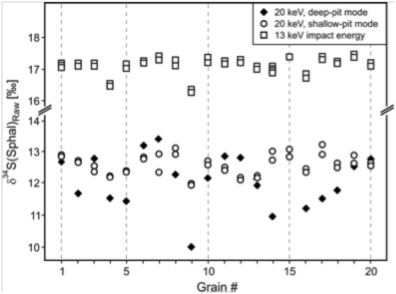
Figure 3. Comparison of raw δ34S values of sphalerite in 20 randomly oriented grains using routine SIMS analytical conditions at 20 keV (black diamonds), a Köhler illuminated beam at 20 keV (white circles), and reduced total impact energy of 13 keV (white squares). Grain-to-grain precision improves from ± 1.7‰ (2SD) for routine SIMS analysis at 20 keV to ± 0.6‰ at 13 keV. The difference in raw values measured at 20 keV and 13 keV results from a change in instrumental mass bias at these different analytical conditions (Kozdon et al. 2010a).

Figure 4. The incident Cs+ beam angle of the SIMS from normal to the sample surface (X-axis) is plotted against the precision in measured δ18O values (Y-axis) for SIMS analyses of magnetite (closed symbols) and hematite (open symbols). The incident Cs+ beam angle (θ) varied from 26 to 14° and a power law fit to these data shows a strong correlation (R2=0.97). Similarly, analyses of magnetite at 20 keV in this study (closed squares) and by Lyon et al. (1998) scaled from 18 to 20 keV (checked squares) show a good correlation (R2=0.72). Spot-to-spot reproducibility for individual grains of magnetite and hematite of ±0.4‰ and ±0.3‰ respectively is plotted at 20 keV (diamonds) and 13 keV (triangles) for reference with no crystal orientation effects. Reducing both the total impact energy and the incident Cs+ beam angle significantly improves the precision in measured δ18O (inset, Huberty et al., 2010b).
-
PROJECT INVESTIGATORS:
-
PROJECT MEMBERS:
Brian Beard
Co-Investigator
Noriko Kita
Co-Investigator
Huifang Xu
Co-Investigator
Philipp Heck
Postdoc
Kenneth Williford
Postdoc
John Fournelle
Research Staff
Jim Kern
Research Staff
Hiromi Konishi
Research Staff
Reinhard Kozdon
Research Staff
Mike Spicuzza
Research Staff
Takayuki Ushikubo
Research Staff
Jason Huberty
Graduate Student
-
RELATED OBJECTIVES:
Objective 4.1
Earth's early biosphere.
Objective 7.1
Biosignatures to be sought in Solar System materials
